Light sheet fluorescence microscopy
We always want to go faster - we just got a new LSFM from 3i. one of the first image sets was 2TB. Those poor IT guys.
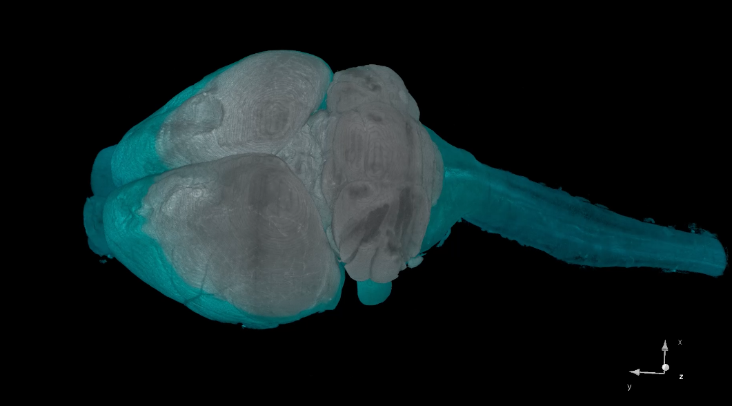
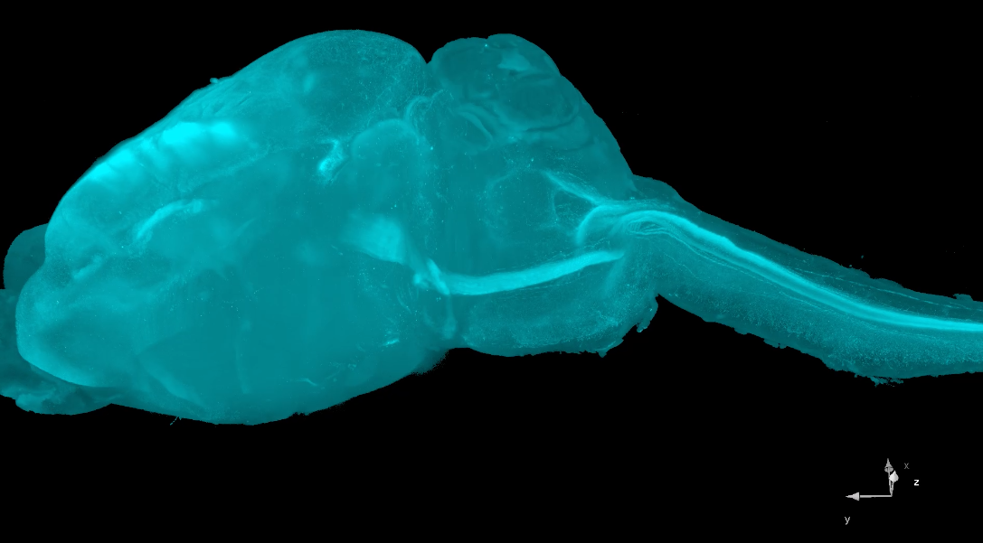
Brain preparation: Kar Men Mah and Abdiel Badillo-Martinez. iDisco by Sensei Pantelis Tsoulfas. Imaging and video by Ed Lachica at 3I
Pantelis Tsoulfas, Jae Lee, Kevin Park and their lab members do most of the work. Vance and John cheer.
3D Visualization of Individual Regenerating Retinal Ganglion Cell Axons Reveals Surprisingly Complex Growth Paths. Bray ER, Noga M, Thakor K, Wang Y, Lemmon VP, Park KK, Tsoulfas P. eNeuro. 2017 Aug 29;4(4). pii: ENEURO.0093-17.2017. doi: 10.1523/ENEURO.0093-17.2017. eCollection 2017 Jul-Aug. PMID: 28856242
Luo X, Salgueiro Y, Beckerman SR, Lemmon VP, Tsoulfas P, Park KK. Three-dimensional evaluation of retinal ganglion cell axon regeneration and pathfinding in whole mouse tissue after injury. Exp Neurol. 2013 Sep;247:653-62.
Soderblom C, Luo X, Blumenthal E, Bray E, Lyapichev K, Ramos J, Krishnan V, Lai-Hsu C, Park KK, Tsoulfas P, Lee JK. Perivascular fibroblasts form the fibrotic scar after contusive spinal cord injury. J Neurosci. 2013 Aug 21;33(34):13882-7.
Soderblom C, Lee D, Dawood A, Carballosa M, Santamaria AJ, Benavides FD, Jergova S, Grumbles RM, Thomas CK, Kevin K. Park KK, Guest JD, Lemmon VP, Jae K. Lee JK, Tsoulfas P 3D Imaging of Axons in Transparent Spinal Cords from Rodents and Nonhuman Primates eNeuro March/April 2015, 2(2) e0001-15.2015 1–24
| experimental_neurology_2013_luo.pdf |